Although tiny, the 1 cubic millimeter of tissue was big enough to contain 57,000 cells, 230 millimeters of blood vessels, and 150 million synapses.
“It was less than a grain of rice, but we began to cut it and look at it, and it was really beautiful,” he said. “But as we were accumulating the data, I realized that we just had way, way more than we could handle.”
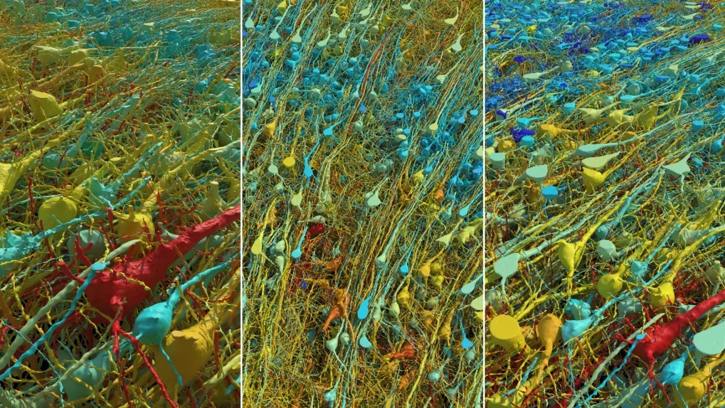
Eventually, Lichtman and his team ended up with 1,400 terabytes of data from the sample — roughly the content of over 1 billion books. Now, after the lab team’s decade of collaboration with scientists at Google, that data has turned into the most detailed map of a human brain sample ever created. This image displays a single human neuron (white) and all of the axons from other neurons that connect to it. The blue threads are inhibitory axons, while the green ones are excitatory axons. Neurons are the cellular building blocks of the nervous system. /Google Research & Lichtman Lab/Harvard University
The brain sample came from a patient with severe epilepsy. Lichtman said it is standard to remove a small portion of the brain to stop the seizures and then analyze the tissue to ensure it’s normal. The sections were cut into thin slices, stained with heavy metals, and imaged using electron microscopy.
The team used AI-based processing and analysis to create a 3D model of the brain tissue. The result is an interactive 3D model of the brain tissue, and the largest dataset ever made at this resolution of a human brain structure. Google made it available online as “Neuroglancer,” and a study was published in the journal Science at the same time, with Lichtman and Jain among the coauthors.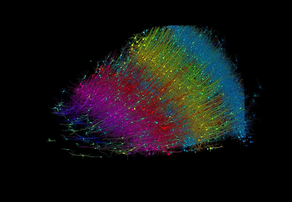 The 3D image above shows excitatory neurons colored by their depth from the surface of the brain. Blue neurons are those closest to the surface, and fuchsia marks the innermost layer.
Lichtman believes that the dataset may contain many more undiscovered details: “That’s why we’re sharing it online, so anyone can look at it and find things,” he added. Next, the team aims to create a full map of a mouse brain, requiring between 500 and 1,000 times the amount of data of the human brain sample.
Mapping an entire human brain would be another 1,000 times bigger, amounting to 1 zettabyte of data, equivalent to the entire internet traffic for the year 2016.